View the
Arrhythmia Recognition Webcast Series
|
 |
Practice ECG 30
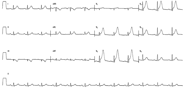
This is a rare occurrence, but one with which you should be intimately familiar -a hyperacute AMI. It occurs during the initial period of the AMI and is a transient finding. It is rare because patients rarely present that quickly to a doctor. Note how tall the T waves appear. They are symmetrical, tall, and broad. The patient will usually have chest pain or pressure, or an anginal equivalent such as dyspnea, diaphoresis, or an indigestion feeling. This patient was having a hyperacute anteroseptal AMI with lateral extension. Look at III and aVF; there are slight depressions in the ST segments consistent with reciprocal changes. The differential diagnosis of these T waves includes intracranial hemorrhage and hyperkalemia. Keep these in mind. The transition in the precordial leads occurs early and the Z axis lies about 10 degrees anteriorly. The prominent R wave is caused by RVH in this case, not a posterior wall AMI. A PWMI would present with ST depressions and inverted T waves rather than ST elevations and hyperacute, positive T waves.
« Back to All
|